

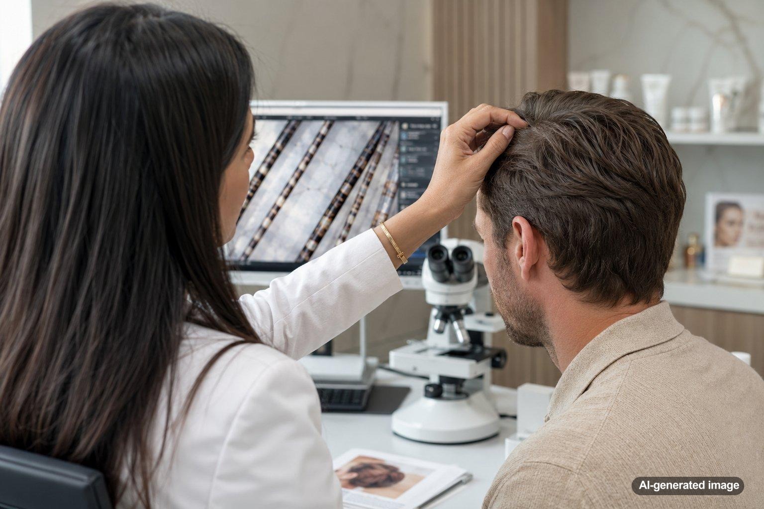
Seeing alternating light and dark sections along a strand of hair under a microscope can be surprising. Some people describe the pattern as a tiger tail, striped hair, or banded hair. The appearance does not automatically point to one condition. It may come from the microscope’s lighting, the way pigment is distributed, residue on the strand, physical damage, or a structural change within the hair shaft.
A dermatologist may examine several hairs rather than relying on one strand. The shape of each band, whether the shaft narrows or folds, and whether the hair breaks at those points can help distinguish a harmless visual effect from a hair shaft abnormality that deserves further evaluation.
Quick answer
- A tiger-tail pattern is a visual description, not a formal diagnosis.
- Regular light and dark bands may reflect pigment distribution or polarized-light effects.
- Nodes, constrictions, fractures, or cup-shaped overlaps can suggest a structural hair shaft disorder.
- Hair dye, heat, chemical processing, traction, and surface residue can alter a strand’s microscopic appearance.
- A dermatologist can compare scalp hairs, eyebrow hairs, symptoms, and medical history before deciding whether testing is appropriate.
What does a tiger-tail pattern actually mean?
There is no single medical finding officially named a tiger-tail hair pattern. The phrase is usually used informally when a strand appears to have repeating stripes, bands, bulges, or alternating thick and thin sections under magnification.
The significance depends on what creates the pattern. A strand may look striped because light passes through areas of different thickness or pigment density. Under polarized light, certain structural properties can also produce bright and dark bands. In other cases, the apparent stripes are separate defects along a fragile hair shaft.
Magnification level matters as well. A low-cost microscope, phone attachment, or uneven light source may create reflections that resemble internal bands. Rotating the hair or changing the lighting can help determine whether the pattern is truly part of the strand.
Common explanations for a banded appearance
Lighting and microscope artifacts
Hair is curved, translucent, and reflective. Light may appear brighter along one edge and darker through the center. Air bubbles, mounting fluid, dust, or compression beneath a slide can add repeating shadows that are not hair disease.
Natural pigment variation
Melanin is not always distributed with perfect uniformity throughout every strand. Gray transition, sun exposure, previous coloring, and normal differences between hairs can create areas that look lighter or darker under magnification.
Cosmetic and environmental damage
Bleaching, permanent color, chemical straightening, frequent high heat, tight hairstyles, aggressive brushing, and repeated friction can weaken the cuticle and cortex. A damaged strand may show cracks, fraying, flattened sections, or irregular nodes rather than a smooth cylinder.
Surface buildup
Conditioner, styling products, dry shampoo, fibers, mineral deposits, and scale from the scalp can cling to a strand. These materials may look like pale bands or sleeves when viewed without professional preparation.
A structural hair shaft abnormality
Some inherited or acquired hair shaft disorders create recognizable patterns of twisting, beading, narrowing, splitting, or telescoping. These findings are uncommon, and their interpretation depends on the exact shape of the defect rather than color alone.
Could it be bamboo hair?
One specific abnormality that can look segmented under magnification is trichorrhexis invaginata, commonly called bamboo hair. In an affected strand, one portion of the shaft folds or telescopes into an adjacent portion. This produces a ball-and-cup or bamboo-node appearance and can make the hair fragile.
Trichorrhexis invaginata is strongly associated with Netherton syndrome, a rare inherited condition that can also involve red or scaly skin, allergic tendencies, and brittle or sparse hair. Finding an unusual pattern in a home microscope image does not establish this diagnosis. The characteristic overlap must be distinguished from reflections, ordinary breakage, and other shaft defects by an experienced clinician.
In some cases, eyebrow or eyelash hairs show a structural change more clearly than scalp hairs. A dermatologist may use trichoscopy, light microscopy, clinical examination, and, when appropriate, additional testing to investigate the finding.
How is it different from other hair shaft changes?
Several abnormalities can create a patterned appearance:
- Trichorrhexis nodosa: Weak points develop along the shaft and may resemble two brushes pushed together. It can occur after repeated physical or chemical trauma and may also accompany certain medical conditions.
- Monilethrix: The strand has regularly spaced thicker and thinner sections that resemble beads on a string. Hair commonly breaks at the narrow points.
- Pili torti: The shaft is flattened and twists along its length, producing alternating bright and dark areas as light reflects from different surfaces.
- Pili annulati: Air-filled spaces within the shaft can produce a banded or speckled appearance, sometimes visible to the naked eye.
- Weathering: Heat, chemicals, ultraviolet exposure, brushing, and friction can create irregular splits and fractures without a repeating inherited pattern.
Because these changes can resemble one another in a photograph, a clear image of one strand is usually not enough for a reliable conclusion.
What you can do at home
Until the pattern is evaluated, gentle hair care can reduce avoidable breakage:
- Pause bleaching, chemical straightening, and other intensive processing.
- Use lower heat settings and keep hot tools moving rather than holding them in one place.
- Avoid tight braids, extensions, ponytails, or clips that repeatedly pull the same area.
- Detangle slowly with conditioner and a wide-tooth comb.
- Reduce friction from rough towels and vigorous brushing.
- Photograph any scalp redness, scale, broken patches, or changes in the eyebrows and eyelashes.
- Bring the microscope images and several naturally shed or broken hairs to your appointment if requested.
Do not rely on supplements or intensive scalp treatments based only on a microscopic image. Hair breakage and shedding can have different causes, and unnecessary products may irritate the scalp or delay a useful evaluation.
What a professional evaluation may include
A dermatologist may begin by asking when the breakage started, whether it has been present since childhood, which hair practices you use, and whether there are associated skin, allergy, or scalp symptoms. The examination may include the scalp, hair density, broken strands, eyebrows, eyelashes, and nails.
Trichoscopy provides magnified views without removing hair. Selected strands may also be examined under light or polarized microscopy. If the pattern raises concern for an inherited condition, the dermatologist may discuss further evaluation, coordination with another specialist, or genetic testing. Testing is selected according to the complete clinical picture, not the appearance of one isolated strand.
When to see a dermatologist
Consider scheduling an evaluation when the pattern is accompanied by:
- Persistent hair fragility or strands that break with minimal handling
- Short, sparse, or uneven hair that does not seem to grow normally
- Loss or breakage of eyebrow or eyelash hair
- Patchy thinning, scalp inflammation, pain, crusting, or significant scale
- Red, itchy, or recurrently scaly skin elsewhere on the body
- A personal or family history of unusual hair fragility
- Symptoms that began in infancy or childhood
Prompt evaluation is especially important for a child with fragile hair plus widespread redness, scaling, recurrent infections, feeding concerns, poor growth, or significant allergy symptoms.
Frequently asked questions
Does a striped hair shaft mean my hair is unhealthy?
Not necessarily. Reflections, pigment differences, mounting materials, and microscope settings can create stripes. The finding is more meaningful when the shaft also shows repeated narrowing, twisting, fractures, or overlapping segments.
Can hair dye create a tiger-tail pattern?
Coloring and bleaching can produce uneven pigment and structural damage, particularly where previous treatments overlap. A clinician can look for cuticle loss, fractures, and other evidence of cosmetic weathering.
Is bamboo hair visible without a microscope?
The individual shaft defect generally requires magnification. However, affected hair may appear short, brittle, sparse, dull, or slow to gain length because it breaks easily.
Can a home microscope identify a hair shaft disorder?
A home microscope may capture a useful clue, but image quality, lighting, magnification, and sample preparation can make normal strands appear abnormal. Professional interpretation usually requires multiple hairs and correlation with an examination.
Should I collect hairs before an appointment?
You may save a few naturally shed or broken hairs in a clean paper envelope, but avoid pulling out a large sample. Contact the office first because the clinician may prefer to select hairs during the examination.
Can damaged hair return to normal?
The visible portion of a damaged strand cannot repair itself biologically. Gentler care may reduce additional breakage while new hair grows, but the outlook depends on whether the cause is cosmetic damage, a scalp condition, or an inherited shaft abnormality.
Ready to get help?
Schedule an appointment or send a message and our team will get back to you.
Prefer to call? 954-666-3736
This article is for educational purposes and is not medical advice. For diagnosis and personalized treatment, please book an appointment with a board-certified dermatologist.
