

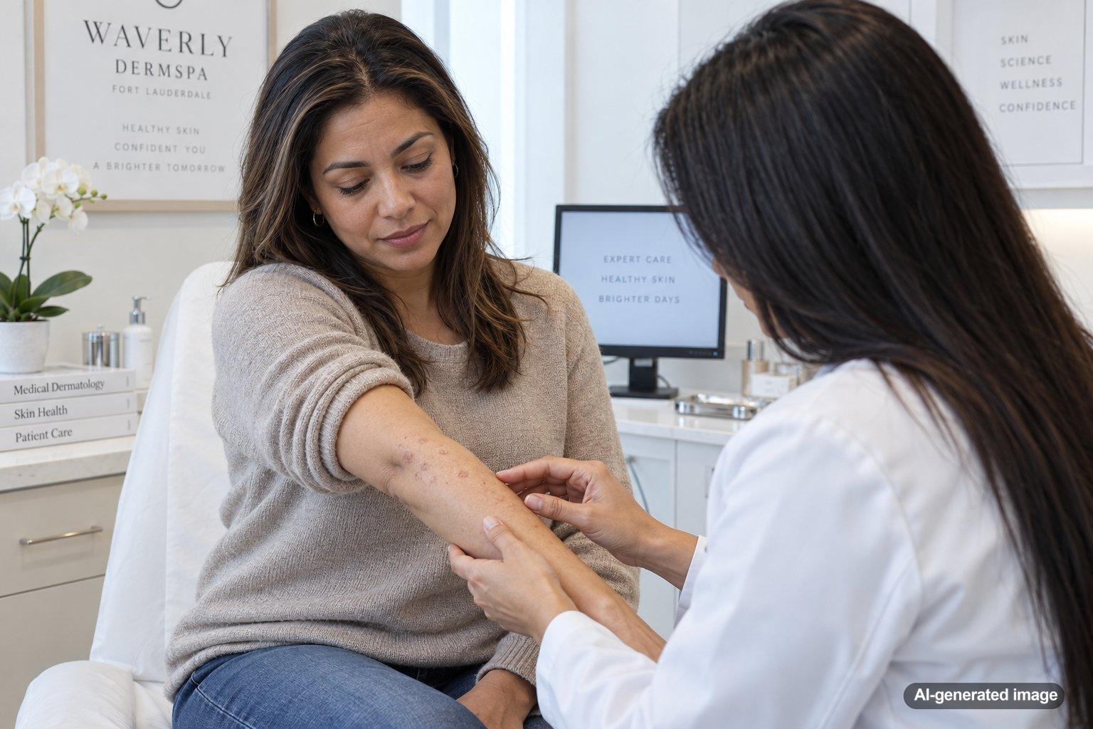
Pityriasis lichenoides is an uncommon inflammatory skin condition that can appear as repeated crops of small red, reddish-brown, or scaly spots. Some eruptions develop gradually and remain relatively mild, while others appear more suddenly and may crust, blister, or become irritated. Because several other skin conditions can look similar, an in-person dermatology evaluation is often important rather than trying to identify the rash from appearance alone.
The condition exists along a spectrum that includes pityriasis lichenoides chronica, often shortened to PLC, and the more acute pityriasis lichenoides et varioliformis acuta, or PLEVA. A dermatologist can evaluate the pattern, symptoms, and evolution of the spots and determine whether additional testing is appropriate.
Quick answer
- Pityriasis lichenoides often causes crops of small red or red-brown bumps that may develop fine scale.
- PLC usually develops more gradually, while PLEVA tends to appear more abruptly and can crust, blister, or erode.
- The exact cause is not fully understood, and proposed triggers include immune reactions associated with infections or certain medications.
- A skin biopsy is sometimes used to help confirm the diagnosis and distinguish it from other rashes.
- Treatment depends on the form, severity, symptoms, and extent of the eruption.
What is pityriasis lichenoides?
Pityriasis lichenoides is a spectrum of inflammatory skin eruptions. The two commonly described ends of that spectrum are PLC and PLEVA, although some people have overlapping features.
PLC generally develops more slowly. It can cause numerous small reddish or reddish-brown bumps, often with a thin, flaky or mica-like scale. New spots may appear while older ones are fading, so lesions at several stages can be visible at the same time.
PLEVA tends to begin more abruptly. The bumps may become crusted, blistered, eroded, or ulcerated in more pronounced cases. Itching or a burning sensation can occur. After individual spots resolve, temporary lighter or darker discoloration may remain, particularly in skin that is prone to pigment changes.
Why can the spots appear in crops?
One recognizable feature of pityriasis lichenoides is that lesions can emerge in waves. Instead of every spot beginning and resolving together, fresh bumps may appear while earlier ones flatten, scale, crust, or leave temporary color changes. This can create the impression of several different rashes occurring at once.
The exact cause of pityriasis lichenoides remains uncertain. Medical literature has proposed several possibilities, including an unusual inflammatory or immune response associated with certain infections, medications, or other immune stimuli. These associations do not mean that a particular infection or medication is responsible in every person.
What can pityriasis lichenoides look like?
The appearance can vary considerably, which is one reason professional evaluation matters. Depending on where a person falls along the PLC-PLEVA spectrum, features may include:
- Small pink, red, or reddish-brown bumps
- Fine or flaky scale over established spots
- Lesions appearing in repeated crops
- Spots at different stages of healing
- Itching, tenderness, or burning in some cases
- Crusting, blistering, or shallow erosions in more acute eruptions
- Lighter or darker marks after inflammation settles
The trunk and areas closer to the shoulders and hips are common sites, although other areas can be involved. The color and visibility of inflammation can also differ across skin tones.
Why a dermatologist may recommend a skin biopsy
Many rashes can produce small scaly or reddish-brown spots. Conditions such as pityriasis rosea, guttate psoriasis, certain medication reactions, insect-bite reactions, and other inflammatory eruptions may enter the differential diagnosis. Less common conditions can also resemble pityriasis lichenoides.
A dermatologist may be able to narrow the possibilities through the history and skin examination, but a skin biopsy is often useful when pityriasis lichenoides is suspected. During a biopsy, a small sample of affected skin is removed and examined under a microscope. The findings can help support the diagnosis and exclude conditions that require a different approach.
What you can do at home
Home care cannot establish the diagnosis, but gentle skin care may make irritated skin more comfortable while you are arranging an evaluation or following a dermatologist’s treatment plan.
- Use a mild, fragrance-free cleanser rather than aggressively scrubbing the spots.
- Apply a simple moisturizer if the skin feels dry or tight.
- Avoid picking at scale, crusts, or healing lesions, which may increase irritation and discoloration.
- Protect exposed areas from excessive sun, especially when healing spots are leaving pigment changes.
- Take clear photographs when a new crop appears, since the eruption may look different by the time of an appointment.
- Keep a list of medications, supplements, recent illnesses, and approximate dates when the eruption changed.
If a product stings or clearly worsens the eruption, stop using it and discuss the reaction with your clinician.
Professional treatment options
Treatment is individualized because pityriasis lichenoides can range from mild, slowly recurring PLC to a more symptomatic acute eruption. A dermatologist will consider how widespread the rash is, whether lesions are crusting or ulcerating, how much itching or discomfort is present, and whether the diagnosis has been confirmed.
Depending on the situation, professional options may include medications used for their anti-inflammatory effects, treatments to reduce itching, or supervised phototherapy. More extensive, persistent, or severe cases may require other prescription therapies. These decisions are best made after an examination because the benefits, limitations, and safety considerations vary from person to person.
When to see a dermatologist
It’s worth scheduling a dermatology evaluation for a persistent or repeatedly recurring crop of unexplained scaly spots, especially when the eruption is spreading, changing, uncomfortable, or leaving significant pigment changes or scars.
Seek more prompt medical attention if a rash is rapidly spreading, becomes very painful, develops extensive blistering or open sores, involves the eyes or mouth, appears with fever or significant illness, or shows signs of infection such as pus, increasing warmth, swelling, or worsening tenderness. Trouble breathing or swallowing, or swelling of the eyes or lips, requires emergency medical attention.
FAQ
Is pityriasis lichenoides contagious?
Pityriasis lichenoides itself is considered an inflammatory skin disorder rather than a rash that simply spreads from one person to another through ordinary contact. Because the exact cause is uncertain and other contagious rashes can sometimes look similar, an examination is important when the diagnosis has not been established.
What is the difference between PLC and PLEVA?
PLC is generally the slower, more persistent end of the spectrum and often features numerous small scaly red-brown bumps. PLEVA usually has a more sudden onset and can produce more inflamed lesions that crust, blister, or erode. Some people show characteristics of both.
Can pityriasis lichenoides leave dark or light marks?
Yes. Healing inflammation may leave temporary areas of darker or lighter pigmentation. The prominence and duration of these changes vary, and they can be especially noticeable in skin tones that readily develop post-inflammatory pigment changes.
Does pityriasis lichenoides always require treatment?
Not every case follows the same course. A dermatologist can help determine whether observation, symptom management, phototherapy, prescription medication, or another approach is reasonable based on the individual presentation.
Can pityriasis lichenoides come back?
It can have a recurring or waxing-and-waning course, particularly PLC. New crops may appear after earlier lesions have begun to fade, so follow-up can be helpful when the eruption is persistent or changing.
Should every suspected case be biopsied?
Not necessarily. The decision depends on how typical the eruption appears, its severity, the possible alternative diagnoses, and the clinician’s judgment. A skin biopsy is commonly considered because microscopic examination can provide valuable diagnostic information.
Ready to get help?
Schedule an appointment or send a message and our team will get back to you.
Prefer to call? 954-666-3736
This article is for educational purposes and is not medical advice. For diagnosis and personalized treatment, please book an appointment with a board-certified dermatologist.
Sources & further reading
- DermNet – Pityriasis lichenoides
- American Academy of Dermatology (AAD) – Rash 101 in adults: When to seek medical treatment
- MedlinePlus (NIH) – Skin Biopsy: MedlinePlus Medical Test
