

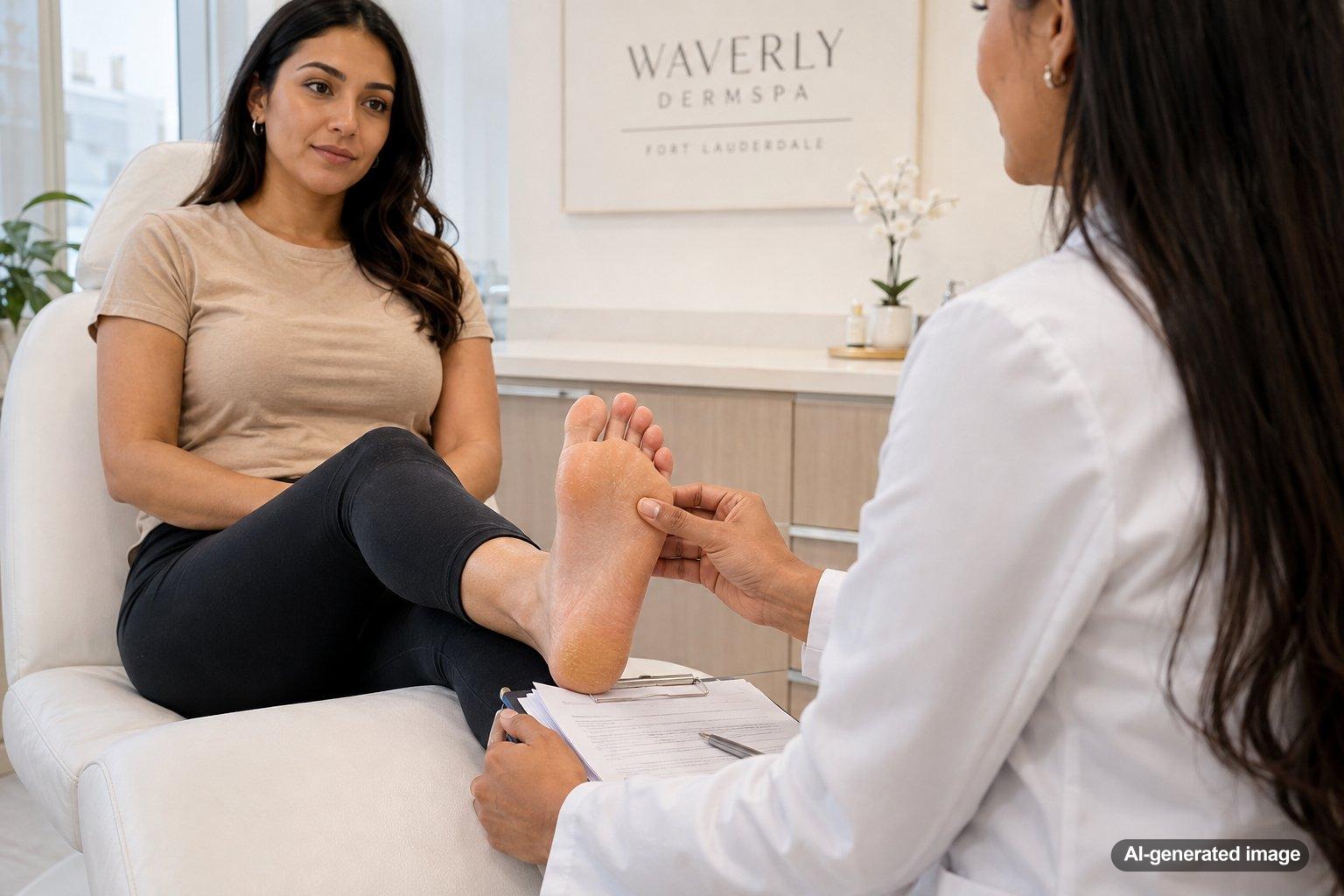
Yellow, thick skin on the soles is often caused by a buildup of keratin, the protective protein that forms the outer layer of the skin. Repeated pressure, friction, dry skin, and footwear can all encourage this layer to become thicker. As compacted skin accumulates, it may take on a yellow, waxy, or slightly translucent appearance.
That appearance is frequently consistent with calluses or another form of hyperkeratosis, but it isn’t possible to identify the cause by color alone. Fungal infection, inflammatory skin conditions, plantar warts, and less common forms of keratoderma can sometimes look similar. The pattern, texture, symptoms, and speed of change help a dermatologist narrow down the possibilities.
Quick answer
- Pressure and friction are common explanations. Thickening often develops beneath the heels, balls of the feet, or other weight-bearing areas.
- The yellow tone may come from compacted keratin. It doesn’t necessarily mean that the skin is stained or infected.
- Scaling or itching may suggest another cause. Athlete’s foot can extend across the soles and may resemble dry, thick skin.
- Location matters. A broad, even layer differs from an isolated rough spot, a wart-like growth, or a changing lesion.
- Persistent or uncomfortable changes deserve evaluation. A dermatologist can examine the skin and determine whether testing is appropriate.
What is causing the yellow, thick appearance?
The outermost skin layer naturally becomes thicker on the soles because the feet must tolerate weight and friction. When pressure, irritation, or inflammation stimulates additional keratin production, the process is called hyperkeratosis. Corns and calluses are familiar examples.
Compacted keratin can look pale yellow, cream, or waxy. It may feel firm and smooth, rough and flaky, or tender when pressure is applied. A callus usually follows a weight-bearing or rubbing pattern, while thickening that covers most of a sole or appears on both palms and soles may require a broader evaluation.
Common causes and possible triggers
- Calluses from pressure or friction: Walking mechanics, prolonged standing, exercise, and shoes that rub or concentrate pressure can lead to firm patches on the heels, forefeet, or toes.
- Dry, cracked skin: Low moisture levels can make thickened skin look rougher and more noticeable. Open-back shoes, frequent barefoot walking, and environmental dryness may contribute.
- Fungal infection: Athlete’s foot may cause itching, burning, peeling, cracking, or scaling between the toes and across the bottoms or sides of the feet. Some patterns produce widespread dry scaling rather than an obvious ring-shaped rash.
- Inflammatory skin conditions: Psoriasis and eczema can affect the soles, creating thick, scaly, itchy, discolored, or cracked areas. Similar appearances can have different causes, so an examination may be needed.
- Palmoplantar keratoderma: This term describes marked thickening of the palms, soles, or both. Some forms are inherited and begin early in life, while others are acquired later and can be associated with skin conditions, medications, repeated exposure, or other health factors.
- Plantar warts: A localized rough growth may become covered by callused skin. Tiny dark dots, interruption of normal skin lines, or tenderness when the area is squeezed can be useful clues, but they aren’t a substitute for an examination.
- Moisture and occlusive footwear: Sweaty feet and shoes that trap heat and moisture can soften the skin, intensify friction, and create an environment in which certain infections are more likely to develop.
What you can do at home
Start with gentle foot care. Wash the feet with a mild cleanser, dry carefully between the toes, and change damp socks promptly. Choose shoes that fit comfortably and allow moisture to escape. Rotating footwear can give each pair time to dry fully.
A fragrance-free moisturizer can help soften dry, intact skin. Products made for thicker foot skin sometimes contain ingredients such as urea or lactic acid, but they may sting cracked or inflamed areas. If you’re unsure which product is appropriate, ask a dermatologist before applying it.
Avoid cutting thick skin with razors, scissors, or other sharp tools. Aggressive filing and strong peeling products can injure healthy tissue beneath the buildup. People with diabetes, reduced circulation, numbness, a history of foot ulcers, or immune suppression should seek professional guidance before attempting to remove callused skin.
If the skin is itchy or scaly, avoid assuming that it is fungal and repeatedly trying different treatments. Conditions such as eczema and psoriasis can resemble athlete’s foot, while untreated fungal infection can persist or spread. Keeping towels, socks, and footwear personal is a sensible precaution while the cause remains uncertain.
Professional evaluation and treatment options
A dermatologist may examine the distribution, skin lines, scale, nails, spaces between the toes, and any isolated growths. When fungal infection is possible, a small skin scraping or another test may help distinguish it from an inflammatory condition. A suspicious or unusual lesion may require closer examination and, in selected cases, a biopsy.
Treatment depends on the cause. Common approaches may include reducing pressure, professionally thinning excess keratin, using moisturizers or keratolytic products, treating confirmed infection, or managing an underlying inflammatory condition. Prescription therapies are selected according to the diagnosis, the condition of the skin, other health factors, and previous treatment response.
If inherited or acquired keratoderma is being considered, a dermatologist may review when the thickening began, whether the palms are involved, family history, medications, and other symptoms. Your clinician can help you decide whether additional evaluation is useful.
When to see a dermatologist
Consider scheduling an evaluation if the thickening is new, widespread, worsening, recurring, or not improving with gentle care. It is also worth getting checked when the cause isn’t clear or when the change affects walking or daily comfort.
- Pain, bleeding, deep cracks, drainage, warmth, swelling, or increasing redness
- Significant itching, burning, peeling, odor, or scaling that extends between the toes
- A single growth with dark dots, an irregular surface, or persistent tenderness
- A sore that doesn’t heal or a spot that changes in color, shape, size, or texture
- Thickening that appears on both the palms and soles or began early in life
- Foot changes in someone with diabetes, poor circulation, reduced sensation, or immune suppression
Rapidly spreading redness, severe pain, fever, or signs of a serious infection warrant prompt medical attention.
Frequently asked questions
Does yellow skin on the soles mean I have a fungal infection?
Not necessarily. Compacted keratin from pressure often looks yellow, but fungal infection can also cause thick scale on the soles. Itching, peeling, involvement between the toes, and nail changes may provide clues. A dermatologist can evaluate the pattern and test the skin when needed.
Why are my heels more yellow than the rest of my feet?
The heels carry substantial pressure and naturally develop a thicker outer layer. Friction, dry skin, standing, exercise, and open-back footwear can make the buildup more visible in that area.
Can I file the thick skin away?
Gentle filing of intact callused skin may be reasonable for some people, but it should never cause pain, bleeding, or raw skin. Avoid sharp tools and aggressive devices. If you have diabetes, poor circulation, numbness, or uncertainty about the diagnosis, get professional guidance first.
What if both soles are thick and yellow?
Symmetrical thickening may still relate to pressure or footwear, but it can also occur with widespread fungal infection, inflammatory disease, or palmoplantar keratoderma. Involvement of the palms, a family history, or onset during childhood makes a dermatology evaluation especially useful.
Can thick skin come back after it is removed?
Yes. If ongoing pressure, friction, dryness, inflammation, or another underlying cause remains, excess keratin may accumulate again. Identifying and addressing the trigger is usually more helpful than repeatedly removing the surface buildup alone.
Ready to get help?
Schedule an appointment or send a message and our team will get back to you.
Prefer to call? 954-666-3736
This article is for educational purposes and is not medical advice. For diagnosis and personalized treatment, please book an appointment with a board-certified dermatologist.
Sources & further reading
- Cleveland Clinic – Hyperkeratosis: What It Is, Types, Causes & Treatment
- American Academy of Dermatology (AAD) – Ringworm: Signs and symptoms
- DermNet – Palmoplantar keratoderma
